Posted on

Axolotl Genetics, Part 3: Melanism and Axanthicism

In part 2 of this article, we went over albinism, a recessive genetic mutation which affects one type of pigment cells (melanophores, responsible for the dark pigment eumelanin). As you can easily guess, there are also mutations which affect the other two types of pigment cells: iridophores, which produce shiny white crystallized purines, and xanthophores, which produce yellow pteridines. In this section, we will focus on these two mutations, which are a bit more complex than albinism.

Melanism

Melanism is a recessive mutation similar to albinism, but instead of affecting melanophores, the mutation acts on iridophores. All axolotls receive either the M or m allele from each parent, which means their genotype for the melanism trait is either:

  • M/M (homozygous dominant)
  • M/m (heterozygous)
  • m/m (homozygous recessive)

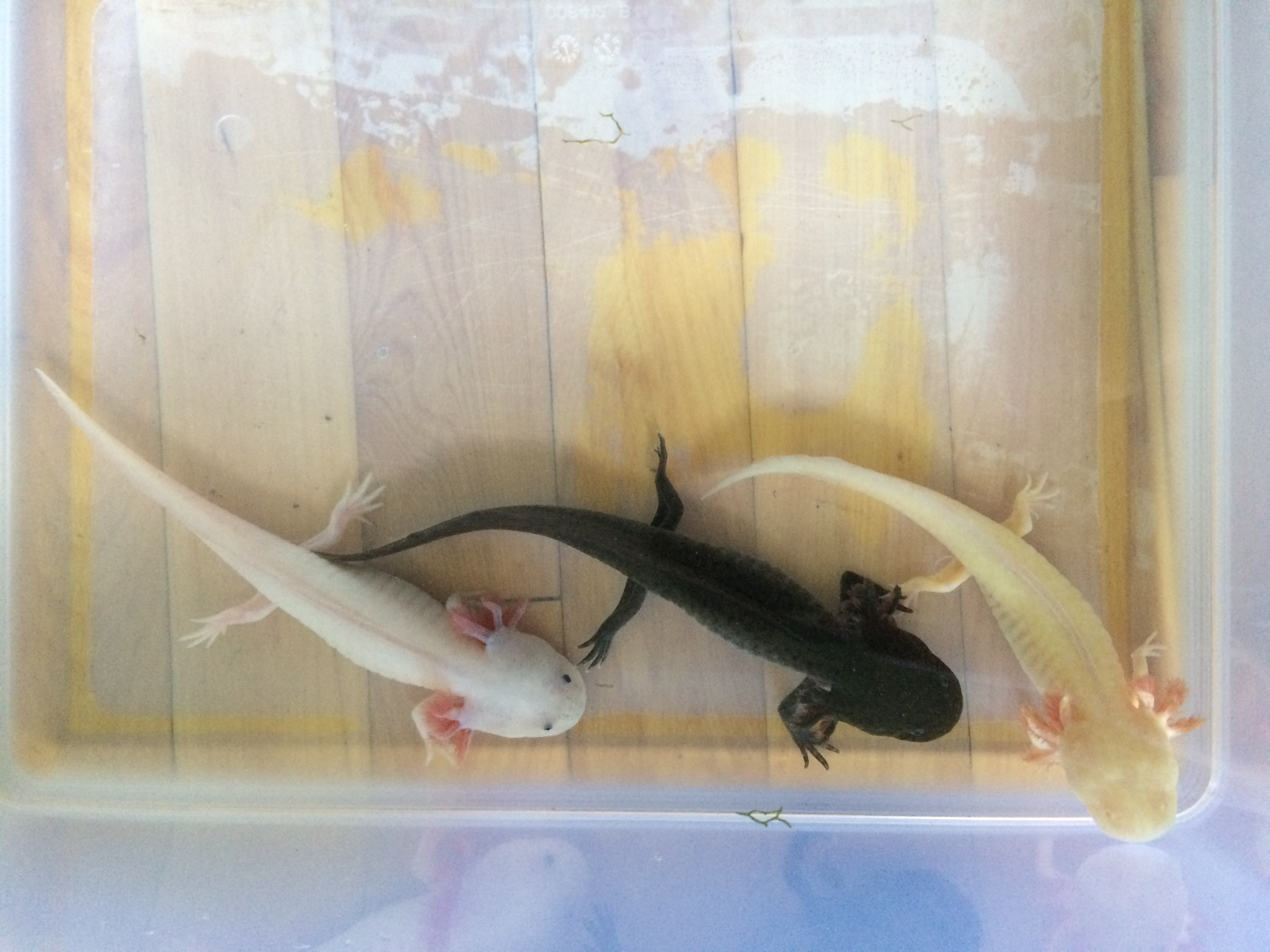
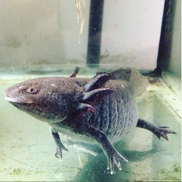
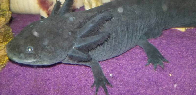
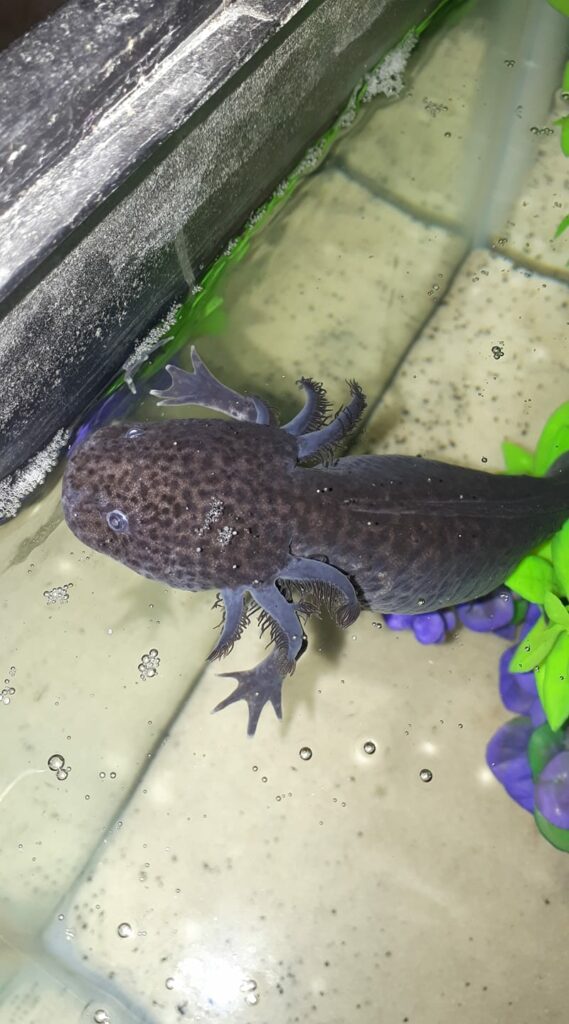
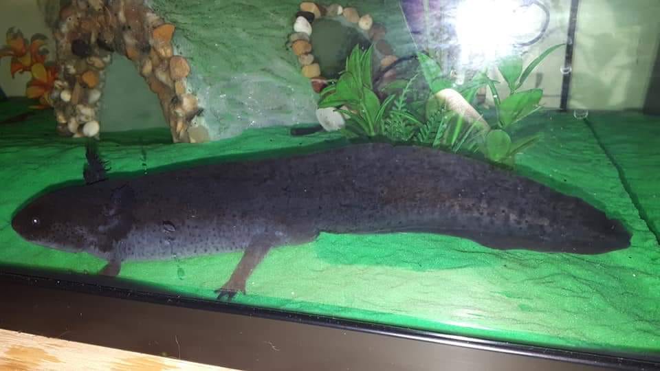
Homozygous dominant and heterozygous axolotls develop normal iridophores, which means they are able to produce crystallized purines (the shiny white pigment). Homozygous recessive axolotls are called melanoids. Since they have no iridophores, they are unable to produce cystallized purines.

This mutation also has a spillover effect: the lack of iridophores triggers the conversion of some xanthophores into melanophores. This is why melanoid axolotls show more eumelanin (black) than any other color morph, and almost no pteridines (yellow). This gives them a grey appearance, which can border on blueish under the right wavelengths.

Due to the reduced number of pteridines, which are important to immune function, melanoid axolotl larvae have a slightly lower survival rate than wild-type or albino axolotls. This is why melanoid axolotls they tend to be a bit more expensive and slightly less common on the market than other color morphs.
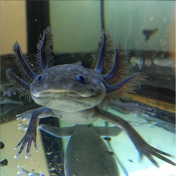
The lower amount of pteridines makes melanoid axolotls appear almost blue under certain wavelengths. Here is X, one of my melanoids, under blue and white LED lights.
This melanoid axolotl has just a hint of yellow pigment on his face, which gives him an almost wild-type look. We can tell that he is a melanoid because his eye ring lacks the metallic shine of wild-type axolotls. Photo by Patricia’s Gill Babies.

This axolotl is both melanoid and albino, which means it has a lower than normal amount of xanthophores (caused by melanism) in addition to a complete absence of melanophores (caused by albinism). This explains why so few yellow pigments are visible on its skin. Photo by Samantha Nicole.

Axanthicism

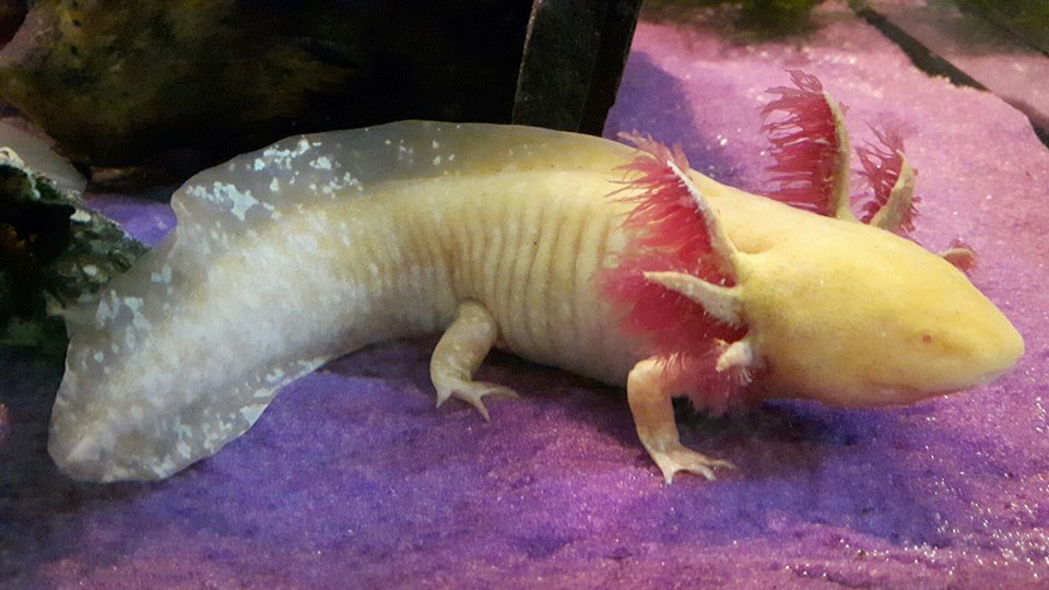
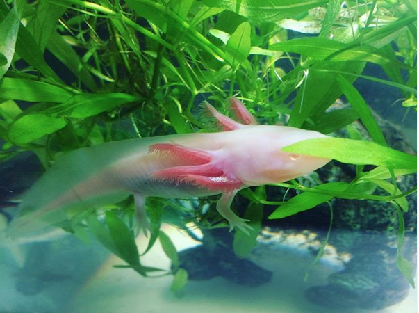
As you can imagine, axanthicism acts on xanthophores, the pigment cells responsible for producing pteridines. But the name of the trait, which means “lack of xanthophores”, is actually misleading. As it turns out, axanthic axolotls do have a certain amount of xanthophores, but those xanthophores are unable to produce pteridines due to a genetic mutation, which is believed to have originated from a virus.

Even though they can’t produce pteridines, the mutant xanthophores are able to store some yellow pigments from the axolotl’s diet (chiefly riboflavin, also known as vitamin B2). This helps compensate a bit for the lack of pteridines, but since they are slowly accumulated over time, axanthic larvae still have a low survival rate compared to other color morphs. This, along with the strict import laws currently in place, explains why axanthic axolotls are nearly impossible to find on the Canadian market.

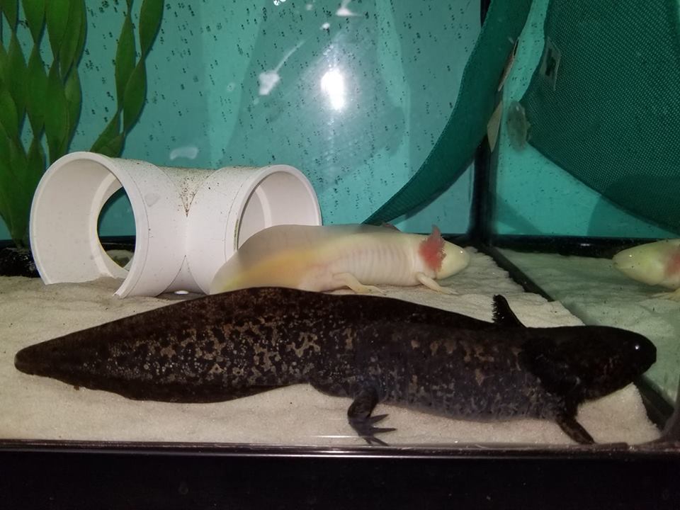
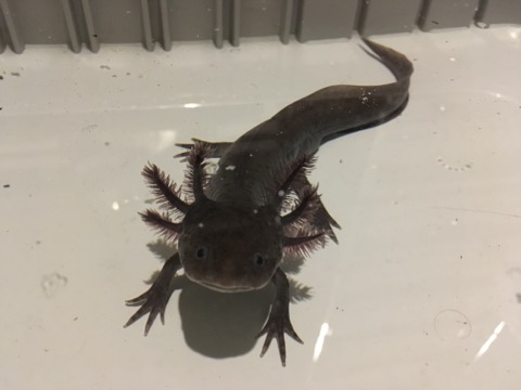
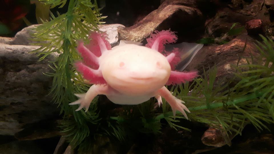
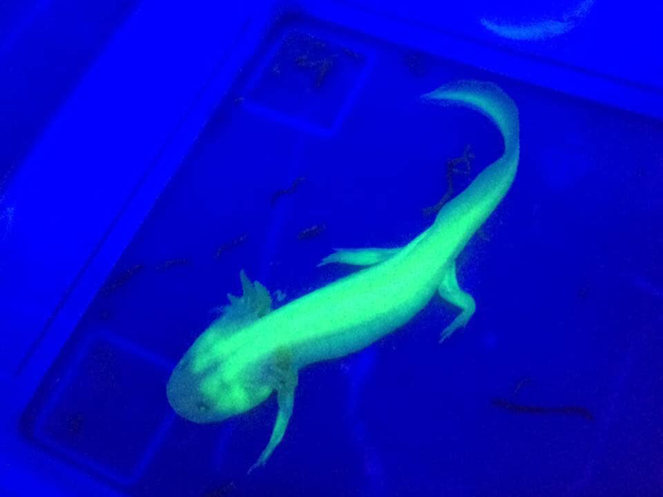
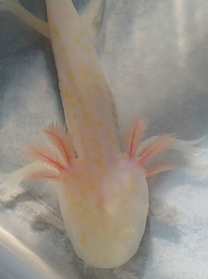
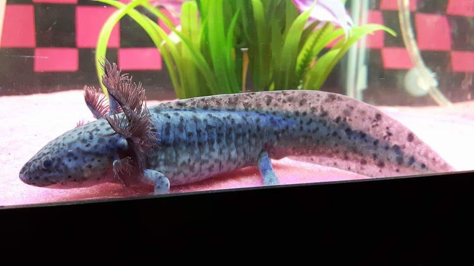
In addition to causing a complete lack of pteridines, the axanthic mutation prevents iridophores from differenciating during development. As a result, axanthic axolotls often look a lot like melanoids. One way to tell them apart is to look at them under a blueish light. The complete absence of yellow pigments at birth tends to give axanthic a purple hue, whereas melanoids are more of a blueish grey. The purple effect tends to fade over time due to the accumulation of other yellow pigments, but some axolotls (such as Sarah, below) do manage to retain it through adulthood.

To make matters more confusing, axolotls can be both axanthic and melanoid. If an axanthic axolotl is especially dark, chances are it is also melanoid, but there is no way to be certain unless the genotype of both parents is known. If an axanthic axolotl accumulates a lot of yellow pigment over the years, then it probably isn’t a melanoid, as melanism further reduces the overall number of xanthophores.

Sarah, showing the purple-grey color characteristic of axanthic axolotls. Photo by Leslee Anne Vanden Top (Axolotl Heaven).
Pale axanthic axolotls such as this one are sometimes called “lavender”. Despite looking similar to light melanoids, lavender axolotls are unlikely to possess the melanism trait. Photo by Leslee Anne Vanden Top (Axolotl Heaven).

Chances are this very dark axanthic male is also homozygous for melanism. Photo by Leslee Anne Vanden Top (Axolotl Heaven).

<- Axolotl Genetics, Part 2: Mendelian Inheritance and Albinism | Axolotl Genetics, Part 4: Leucism, Copper and GFP [Coming Soon!]

Posted on

Axolotl Genetics, Part 1: Color Pigments

An axolotl’s coloring is the result of genetics, and to a lesser degree, environment and diet. Let’s go over the different color pigments involved, and you’ll understand what I mean.

The three natural color pigments are:

  • Eumelanin (brown, black)
  • Crystalized purines (iridescent white)
  • Pteridines (yellow, orange)

There is also a fourth pigment that is present in some transgenic axolotls:

  • Green fluorescent proteins (bright yellow, glowing neon green under a UV light)

We’ll get back to this one later — let’s focus on the three natural pigments first. These are naturally present in the majority of axolotls. Besides looking pretty and helping with camouflage, they also come with health benefits: eumelanin helps protect the skin against UV radiation, and pteridines play an important role in the axolotl’s immune system.

You can see all three pigments expressed in the picture below:

Two of my light wild-type axolotls, showing all three natural pigments: eumelanin, pteridines and crystallized purines.

Axolotls that possess all three pigments are called wild-type. Even though they all have the same pigments, there can be a lot of variation in wild-type appearance. For instance, the axolotls shown above have a lot of yellow pteridines, which gives them an overall olive tint. They also have white spots on their tails. If I had taken the photo with the flash on, you would have seen that those white spots are shimmery, because they are made of crystallized purines.

The axolotl in the photo below is a much darker wild-type:

Katla, one of my dark wild-type axolotls, showing a predominance of eumelanin.


In this photo, we can see a lot of eumelanin. The other pigments are also present, but not very noticeable. You can see a little bit of crystallized purines in the eye ring and the tip of the gill stalks. Pteridines are almost completely invisible under the dark eumelanin.

Let me show you one more, very different wild-type look:

A “starburst” wild-type axolotl (front). Photo by Patricia’s Gill Babies


Isn’t this boy gorgeous? Here, eumelanin forms the base skin color, but the pteridines and crystallized purines being layered on top of each other create a gold flake effect.

In addition to the variety among wild-types, there are a lot of different color types, or “morphs”, besides wild-type. Over the course of their history, axolotls have undergone several genetic mutations which affect their pigmentation — some of which are natural, some of which are the result of human intervention.

Here are the six main genetic traits that affect axolotl pigmentation:

  • Albinism (affects eumelanin)
  • Melanism (affects crystallized purines)
  • Axanthicism (affects pteridines and crystallized purines)
  • Leucism (affects eumelanin, pteridines and crystallized purines)
  • Copper trait (affects eumelanin and/or pteridines)
  • GFP trait (affects green fluorescent proteins)

We’ll talk more about these traits in the next section of the article. For, now I just want you to keep in mind that there are several genetic traits that can essentially switch pigment production on and off, or affect how pigments are distributed around the body.

Let’s take a closer look at what each pigment looks like individually.

Eumelanin

Eumelanin is the pigment responible for shades of brown and black. It is produced by pigment cells called melanophores. To give you a better idea of what the pigment looks like on its own, here is what an axolotl looks like when it shows only eumelanin:

My melanoid axolotl, Z, showing only the pigment eumelanin.


Fun fact: the amount of eumelanin produced by an axolotl depends on two things: genetics, and environment. Axolotls whose parents were especially dark tend to exhibit similarly dark features. Axolotls who grow up in dark environments also tend to exhibit darker features than ones kept in lighter environments.

The absence of eumelanin, due to an inability to produce melanophores, is called albinism. Here is what an axolotl looks like when you completely remove eumelanin, keeping only the other two pigments:

A golden albino axolotl, showing pteridines and crystallized purines, but no eumelanin. Photo by Patricia’s Gill Babies


Pretty neat, right?

Crystallized purines

Crystallized purines are iridescent white pigments, which means they shimmer in a sort of rainbow effect. Combined with pteridines, they can also create a shiny golden color, as we’ve seen above. Crystallized purines are produced by pigment cells called iridophores. Here is what iridophores look like on their own:

One of my “starlight” white albinos, showing crystallized purines concentrated on the gill stalks and eye ring.


The inability to produce iridophores is called melanism. Notice how the shiny white pigments are missing in the picture below:

A melanoid white albino axolotl, showing a lack of crystallized purines. Photo by Patricia’s Gill Babies


Melanism is a little bit more complex than albinism. We’ll talk about it more in part 3 of this article.

Pteridines

Pteridines are responsible for yellow and orange coloration. They are produced by pigment cells called xantophores. This is what pteridines look like when you remove the other two pigments:

A melanoid golden albino axolotl (juvenile), showing only pteridines. Photo by Samantha Nicole.


The inability to produce pteridines is called axanthicism. Axanthic axolotls are exceedingly rare, if not impossible to find in the Canadian pet trade. This is partly due to strict import laws, and partly due to the effect axanthicism has on axolotl health. Since pteridines play a role in immune function, axanthic axolotls have a lower survival rate than other axolotls.

In the absence of pteridines, axanthic axolotls take on a purple-grey look:

Sarah the axanthic axolotl, showing a lack of pteridines. Photo by Leslee Vanden Top


Do you notice some odd things about this picture? Axanthicism is a much more complex mutation than albinism and melanism. We’ll talk more about it when we get to the next section.

Green fluorescent proteins (GFP)

In the course of their use as animal research models [more on this soon!], some axolotls got a pretty cool addition to their genomes: the GFP trait. Originally found in a species of jellyfish, this trait causes nearly every cell in the axolotl’s body to produce a bright yellow protein which glows neon green under a UV light. Why is this cool? First, it’s been very helpful to researchers working on limb regeneration and organ transplants. Second, it looks very pretty! And third, the trait can be passed down from generation to generation. But my favorite thing about it is that, since the effect isn’t limited to pigment cells, it isn’t affected by leucism. You’ll see what I mean when we get to the next part!

A GFP leucistic axolotl under UV light. Photo by Carey Lynn Cooper.


Now that you have a good idea of what the individual pigments do, let’s take a look at the genetics behind them!

Axolotl Genetics, Part 2: Mendelian Inheritance and Albinism ->